What Is an X-Ray CT Machine?
 An X-ray CT machine is a device that irradiates X-rays to examine the materials and structure inside an object.
There are two main types of X-ray CT systems: medical and industrial.
Medical X-ray CT machines can examine bones, muscles, blood vessels, and other tissues from the head to the feet, as well as internal organs.
Uses of X-Ray CT Machines
There are two main types of X-ray CT machines: medical and industrial. They are explained in the following order.
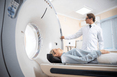
1. Medical X-Ray CT Machines
Medical X-ray CT machines detect X-rays transmitted through the human body to obtain information on the inside of the human body, and are used to determine medical conditions. There are two types of medical X-ray CT Machine: simple CT, which observes the human body as it is, and contrast-enhanced CT, in which a contrast agent is injected into the blood vessels for observation.
2. Industrial X-Ray CT Machines
Industrial X-ray CT machines are widely used in nondestructive inspection of materials, and such industrial X-ray CT machines are called “observation CT”. Examples of nondestructive inspection include shape evaluation and defect investigation of semiconductor packages, internal structure evaluation of tablets, and shape and fiber orientation evaluation of carbon fiber reinforced plastics.
In recent years, many industrial X-ray CT machines have also been developed for measurement purposes. Like CT for observation, CT for measurement enables nondestructive inspection, size measurement, and data analysis using CAD.
The “CT for measurement” enables full acquisition of the three-dimensional shape of the object, high-precision shape measurement, and high-speed inspection and measurement.
Principle of X-Ray CT Machines
There are two types of X-ray CT machine: medical and industrial. Both systems irradiate an object with X-rays from all directions in 360 degrees to examine the transmitted and absorbed X-rays, but the imaging methods used in medical and industrial applications are different.
1. Medical X-Ray CT Machines
A medical X-ray CT machine is characterized by a doughnut-shaped gantry and a bed that moves slowly through the center hole of the gantry while a human body is lying in the center hole of the gantry. Inside the gantry, an X-ray tube that irradiates X-rays and a detector that detects X-rays irradiated from the X-ray tube is placed across the center hole.
When a human body on a bed enters the center hole of the gantry, some of the X-rays emitted from the X-ray tube are absorbed by the human body, and the rest are transmitted and detected by the detector. At this time, the gantry rotates around the bed, irradiating X-rays from 360 degrees and detecting the transmitted X-rays.
The gantry then rotates around the bed, irradiating X-rays from 360 degrees and detecting the X-rays transmitted through the gantry. Helical scan is commonly used in this medical X-ray CT machine.
Helical scan is an imaging method in which the bed moves slowly through the center hole of the gantry while continuously irradiating and detecting X-rays. This produces a series of horizontally sliced images of the human body in the longitudinal direction in the direction of height.
2. Industrial X-Ray CT Machines
Unlike medical X-ray CT machine, industrial X-Ray CT Machine is characterized by the fact that the material under investigation rotates instead of the X-ray tube and detector being fixed. There are two types of industrial X-Ray CT machines: the horizontal irradiation type and the vertical irradiation type.
The horizontal irradiation type has the X-ray tube, material, and detector arranged horizontally, while the vertical irradiation type has them arranged vertically. The appropriate type is selected according to the material to be investigated and the location to be investigated.
Other Information on X-Ray CT Machines
1. Multi-Slice Medical X-Ray CT Machines
Medical X-ray CT machines include multi-slice medical X-ray CT machines with multiple rows of detectors arranged in the direction of bed movement.
In this single-slice X-ray CT machine, the detectors are arranged in the direction perpendicular to the direction of bed movement, i.e., transversely for humans, and are distributed in a single row in the direction of bed movement. Therefore, when the gantry makes one rotation, only one cross-sectional view is constructed.
In contrast, the multi-slice medical X-ray CT machine has multiple rows of detectors distributed in the direction of bed movement, enabling acquisition of multiple cross-sectional views with a single rotation of the gantry. This enables imaging in a short time and reduces the burden on the human body. 3D images can also be constructed by combining this with helical scanning.
2. 3D Image Processing Methods for X-Ray CT Machines
The following three types of 3D image processing methods are used in X-ray CT machine technology.
Multi-Panel Reconstruction (MPR)
Multi-Panel Reconstruction (MPR) is characterized by its ability to construct images from three-dimensional data in the coronal and sagittal cross sections, which are cross sections in the direction of the human height, in addition to the transverse cross section of the human body. This method is the most used in current CT 3D processing.
Maximum Image Projection (MIP)
In the maximum intensity projection (MIP) method, an arbitrary viewpoint is set for the three-dimensional data. The maximum value along the path between the viewpoint and the projection plane is then projected onto a two-dimensional surface.
It is characterized by its ability to minimize the effect of image noise and to output images with good contrast, even if they are low-contrast images. However, since values other than the maximum value are not reflected in the image, several angles of observation are required if the front and rear positions are to be determined correctly.
Volume Rendering (VR)
The volume rendering method (VR) sets the upper and lower limits of CT values for the area of interest. The image is then constructed by adding parameters corresponding to opacity to the set range and performing shading processing. This method is suitable for 3D images of blood vessels, etc.