What Is a Polarizing Microscope?
A polarizing microscope is an optical microscope that selects and observes polarized light.
Optical microscopes used in science experiments observe all light reflected from a material through an eyepiece. Light is a wave in which the electric and magnetic fields oscillate in a direction perpendicular to the direction of travel. Light with a regular direction of oscillation in the electric field is called polarized light. A polarizing microscope observes polarized light that is reflected from a material and vibrates in a specific direction.
Linearly polarized light is shone on a material, and changes in the polarization state can be observed as color or light/darkness. When polarized light is selected and observed with a microscope, it is possible to identify the state and components of a substance.
Uses of Polarizing Microscopes
Polarizing microscopes were originally used to determine the state and composition of minerals. However, they are now also used in the development of polymers and biotechnology. Changes in polarization state reflect molecular orientation and crystal structure, allowing evaluation of the internal structure of polymers. Furthermore, in combination with temperature control equipment, phase transition behavior can be observed.
One of the major discoveries made using polarizing microscopes is liquid crystals. Liquid crystals, which are liquid but have a molecular arrangement similar to that of solids, were first observed with polarizing microscopes, leading to the development of today’s liquid crystal televisions and other products.
In addition, many biological materials have a state and molecular structure equivalent to liquid crystals, and polarizing microscopes will continue to play an active role in the medical and pharmaceutical fields in the future.
Principle of Polarizing Microscopes
Polarizing microscopes use filters to select the polarization of light, producing optical microscope images that reflect the optical properties of the sample.
1. Composition of Polarizing Microscope
An ordinary optical microscope consists of a light source, a sample stage, and an objective lens. Light emitted from the light source strikes a material, which enters the objective lens and can be observed through the eyepiece. The principle of polarizing microscopes is basically the same as that of an optical microscope, except that a polarizer is placed between the light source and the sample, and two polarizing plates called an analyzer (analyzer) are placed between the objective lens and the eyepiece lens.
The light emitted from the light source is natural light, the same as fluorescent light, including all directions. This light is converted to polarized light by transmitting it through the polarizer and shining it on the material to be observed. Polarized light that has changed direction when transmitted through a material passes through an analyzer in a cross-nicol arrangement that is perpendicular to the polarizer and can be observed.
2. Image of Polarizing Microscopes
When a sample with no anisotropic refractive index is observed with polarizing microscopes, the linearly polarized light emitted from the polarizer does not change its polarization state and cannot pass through the analyzer, so the field of view when observed through the eyepiece is dark.
When observing a sample whose refractive index differs according to the direction of polarization (birefringence), if the direction of oscillation of the incident linearly polarized light is aligned with the optical axis of the sample, the polarization state of the incident light does not change and the field of view is dark, as described above. On the other hand, when the oscillation direction of the incident light is different from the optical axis of the sample, the incident light is divided into two polarization components due to the birefringence of the sample, and the composite component is different from the polarization state before the sample transmission. The change in polarization state causes the light to pass through the analyzer, resulting in a bright field of view.
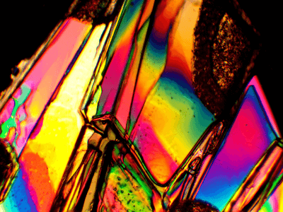
Images from a polarizing microscope appear tinted due to the optical path difference between the two light components caused by the birefringence of the sample. In polarizing microscopes, the stage on which the material is placed can be rotated 360° in order to change the angle of polarized light with respect to the optical axis of the sample.
Other Information on Polarizing Microscopes
Applications of Polarizing Microscopes
Polarizing microscopes use techniques that can be used in combination with other optical measurement methods because they can study crystal domains and even their orientation.
1. Fluorescence Measurement
Polarizing microscopes can be used in combination with fluorescence measurement. Normal fluorescence measurements are ensemble information obtained from various positions and orientations of the crystal domains. However, since the optical properties change depending on the orientation of the crystal domains, polarizing microscopes, which can identify the crystal orientation, come into play. Polarizing microscopes allow us to observe the emission of polarization information in a specific direction by injecting a laser with a specific direction of polarization in the incident light.
2. Time-Resolved Measurement
Polarizing microscopes can also be used in combination with time-resolved spectroscopy. While ordinary time-resolved spectroscopy provides ensemble information from various positions and orientations of crystal domains, polarizing microscopy allows time-resolved spectroscopic measurements of absorption and emission by determining the orientation and position of crystal domains.