What Is a Confocal Microscope?

A confocal microscope is a type of optical microscope designed to eliminate out-of-focus light by incorporating a pinhole at the focal plane in front of the detector. This allows for the capture of sharp, blur-free images.
It achieves three-dimensional, all-in-focus imaging of an object by collecting blur-free images at various depths and reconstructing them.
The confocal microscope, also known as a confocal laser scanning microscope (CLSM) or laser scanning confocal microscope, is renowned for its ability to produce high-resolution three-dimensional images.
Uses of Confocal Microscopes
Confocal microscopes are utilized for measuring and analyzing the shape of objects with high sensitivity and resolution. They are ideal for non-contact measurements using laser technology, especially for soft or delicate objects that could be damaged by contact. The small diameter of the laser beam enables the observation of minute details and irregularities.
These microscopes find applications in industrial inspections and the life sciences for cell and organism observation.
1. Industrial Applications
Confocal microscopy can measure various surface characteristics, such as average height, differences from reference areas, and surface roughness. It generates color three-dimensional images, facilitating intuitive observation and measurement.
3D images enable specific area measurements, allowing for the quantification of scratches and dents and the assessment of product characteristics based on surface area changes.
Inspections differentiate between acceptable and defective products, improving overall work processes. Applications include printed circuit boards and large LCD panel inspections.
2. Life Science Applications
In life sciences, confocal microscopy is used for three-dimensional visualization of cells and tissues, recording changes over time in living specimens to explore and elucidate their functions.
3. Measurement Device Applications
Confocal microscopy principles are applied in various measurement devices, such as Raman microscopes for material identification and multiphoton excitation microscopes for deep tissue observation.
Principle of Confocal Microscopes
1. Structure
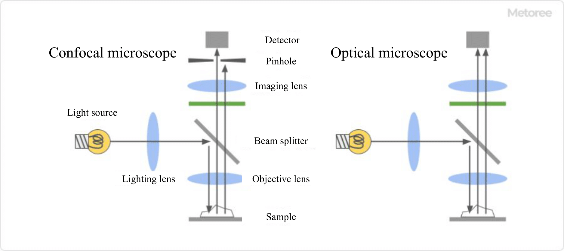
Figure 1: Structure of Confocal Microscope and Optical Microscope
The confocal microscope primarily consists of a laser light source, a beam splitter, lenses, a pinhole, and a detection unit.
Light from the source is directed through the lens to the specimen via the beam splitter. Reflected light from the specimen passes through the lens and beam splitter again, reaching the detector through the pinhole.
2. Operating Principle
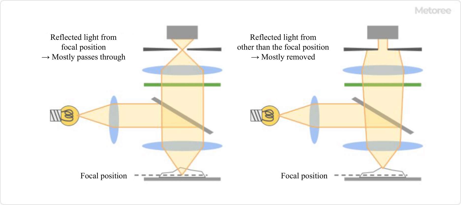
Figure 2: Principle of Confocal Microscope
The confocal microscope scans a focused laser beam across the sample, detecting light that is reflected. Only light from the focal point is detected through the pinhole, enhancing depth resolution and reducing blur compared to traditional optical microscopes.
Types of Confocal Microscopes
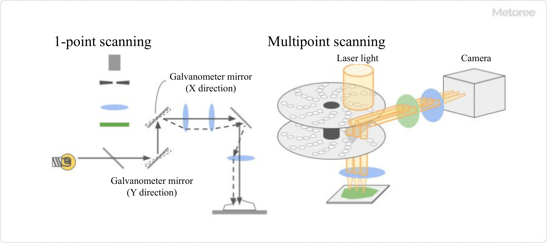
Figure 3. Types of Confocal Microscopes
Confocal microscopes are categorized into single-point and multi-point scanning types.
The single-point type uses a galvanometer scanner for scanning, with a pinhole to eliminate blur. Innovations like acousto-optic deflectors (AODs) and resonant mirrors enhance scanning speed.
The multi-point type employs a rotating disk with multiple pinholes for scanning, with a CCD or CMOS camera detecting the light. This method is faster and suitable for high-speed observations and time-lapse imaging. Line scanning types, using a slit for linear measurements, are also available.