What Is a Fluorescence Microscope?
Fluorescence Microscope is a device to observe the fluorescence of fluorescent substances in an object by using laser light, super high-pressure mercury vapor lamp, or xenon lamp as a light source. In ordinary optical microscopes, visible light such as halogen lamps are used as a light source to irradiate an object and observe reflected light and transmitted light.
Fluorescence Microscope is a type of microscope that mainly targets biological tissues and cells labeled with fluorescent substances. Since the resolution of a microscope depends on the wavelength of the light used, Fluorescence Microscope, which uses light with a short wavelength, is characterized by its superior spatial and temporal resolution.
Therefore, highly quantitative information can be obtained. Fluorescence Microscope is becoming more and more important as its functions are becoming more sophisticated as confocal laser microscopy and multiphoton microscopy.
Applications of Fluorescence Microscope
Fluorescence Microscope is mainly used for bio-imaging. The specific targets are cells and tissues, which can be observed while alive. The following techniques are used in combination to label objects with fluorescence:
- Technology to fluorescently label specific proteins through genetic recombination or other means
- Labeling nucleic acids and other substances with fluorescently labeled chemicals
- Technology to express fluorescent proteins in specific cells
These technologies enable us to observe the localization of target proteins and expressed genes. In addition, drugs and proteins that emit fluorescence in response to specific substances have been developed, making it possible to visualize neural activity and intracellular dynamics of substances.
In recent years, the advent of CRISPR technology has made the creation of genetically modified organisms much easier, and the scope of its application is rapidly expanding.
Principle of Fluorescence Microscope
Fluorescence Microscope is an instrument for observing fluorescence. Fluorescence is emitted when a fluorescent substance absorbs specific light as energy (excitation light) and releases the energy again.
Exposure to excitation light causes rapid emission of light. The wavelength of fluorescence is longer than the wavelength of the excitation light, and these wavelengths vary with the fluorescent material. Fluorescence Microscope has a filter unit to observe specific fluorescence, which consists of:
- A filter that transmits the excitation light from the light source
- A filter that transmits the emitted fluorescence
- A mirror to prevent interference of the excitation light with the fluorescence
By changing or combining filter units, various fluorescent materials can be observed from the same specimen.
Other Information on Fluorescence Microscope
1. Resolution of Fluorescence Microscope

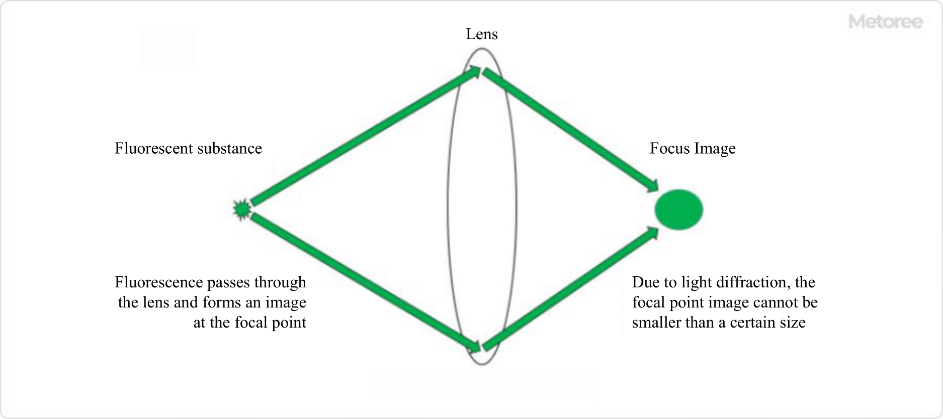
Figure 1. Resolution of fluorescence microscope
The resolution of a microscope refers to “the minimum distance at which two close points can be distinguished from different points. Microscopes use lenses to magnify and observe objects, and in principle, it is possible to increase the magnification infinitely by combining lenses.
However, in the case of optical microscopes, which use light to observe samples, the limit of resolution is approximately half the wavelength of light due to diffraction, which is a characteristic of light. This was considered the theoretical limit of microscope resolution, but a technology was developed that broke through this limit, and the developer was awarded the Nobel Prize in Chemistry in 2014.
The technique is called “super-resolution microscopy. Until the development of super-resolution microscopy, the resolution limit of Fluorescence Microscope was approximately 250 nm, but with super-resolution microscopy, the resolution can be as high as 15-100 nm, which is close to that of electron microscopy. Super-resolution microscopy achieves high resolution by using various techniques to avoid the limiting factors of resolution.
Super-resolution microscopy methods that have dramatically improved resolution and won the Nobel Prize in Chemistry include “PALM” and “STED”. PALM and STED have achieved breakthroughs in Fluorescence Microscope resolution by utilizing special optics and special dyes. Super-resolution microscopes using various other technologies have been produced and are being commercialized by various companies.
2. Advantages of Fluorescence Microscope

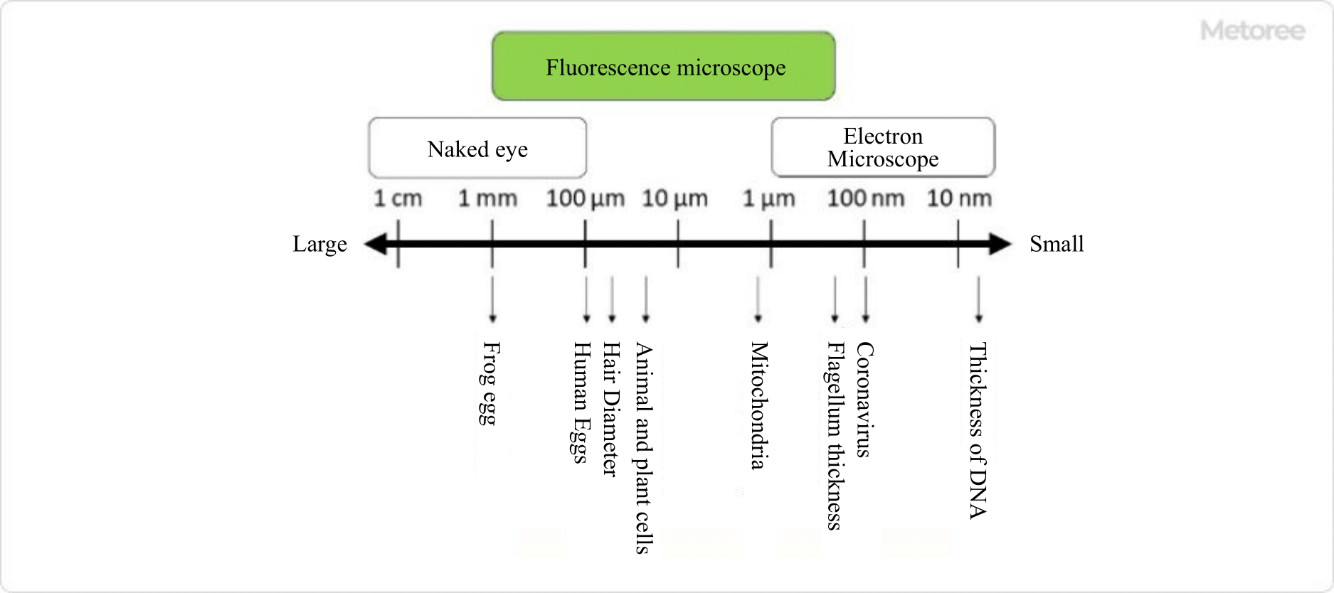
Figure 2. Objects that can be observed with a fluorescence microscope
The advantage of Fluorescence Microscope is that you can observe the behavior of molecules and the structure of cells in detail as visual information. By using the appropriate Fluorescence Microscope for your purpose, you can observe objects with high temporal and spatial resolution.
It is also possible to observe an object using multiple dyes. For example, when two different proteins are labeled with red and green fluorescent substances and observed, a yellow area indicates that these two proteins may exist in the same location in the cell.
A variety of fluorescence materials and Fluorescence Microscopes have been developed for different purposes and applications, and are becoming increasingly important in life science and clinical research.
