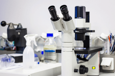
What Is a Phase Contrast Microscope?
 A phase contrast microscope is a type of optical microscope that converts the phase difference of light into contrast for observation.
With an ordinary optical microscope, differences in the reflection and absorption spectra of light from different parts of a sample are observed as differences in brightness or color (contrast). However, when observing nearly colorless materials such as living cells, microorganisms, and bacteria, these contrasts are almost nonexistent, and information such as shape cannot be obtained.
Even if a material is colorless and transparent, if its refractive index differs from that of its surroundings, the diffraction of light will occur at the boundary. Phase contrast microscopes use the phase difference between the diffracted light and the light traveling straight through the material to create a contrast between light and dark, making it possible to observe colorless transparent materials.
Uses of Phase Contrast Microscopes
Phase contrast microscopes are widely used in biology and medicine for observation of cultured cells and clinical examination. Periodontal bacteriological examination in dental clinics is a familiar application for the public. It helps to motivate patients to take better care of their oral health by letting them know how their own oral bacteria are doing.
Phase contrast microscopes allow observation of live cells without the need to stain the specimen. When observing colorless cells with a conventional optical microscope, the sample is stained for observation, but this method has the disadvantage that the staining is time-consuming and kills living cells.
In addition, phase contrast microscopes are also effective in analyzing asbestos, a toxic substance. One method is the dispersion staining method, in which crystals in the immersion solution with a specific refractive index are irradiated with polarized light under a phase contrast microscope, and the color produced is used to determine whether the sample is asbestos or not.
Principle of Phase Contrast Microscopes
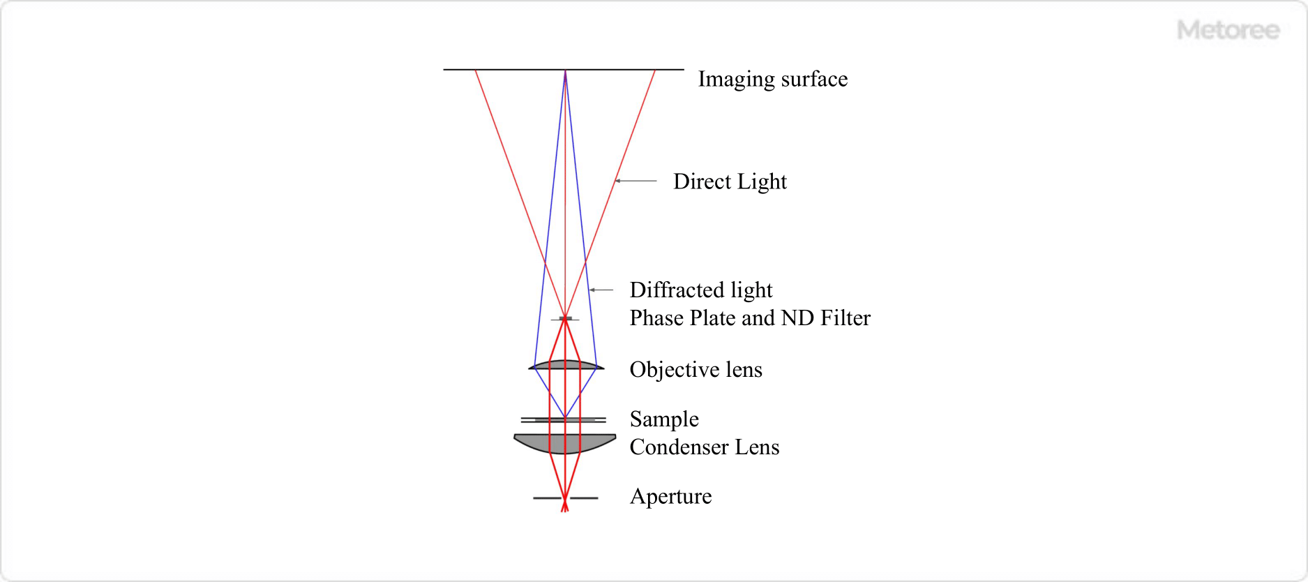
Figure 1. Optical system of a phase contrast microscope
In a phase contrast microscope, a phase plate is inserted only at the position where the direct light passes between the objective lens and the image plane to advance or retard the phase of the direct light by 1/4λ. At the same time, a ring-shaped ND filter is inserted to reduce the intensity of the direct light, but does not change the phase or brightness of the diffracted light.
By these operations, the phase difference between the direct light and diffracted light becomes 1/2λ or 0, and the light and dark contrasts are created by interference.
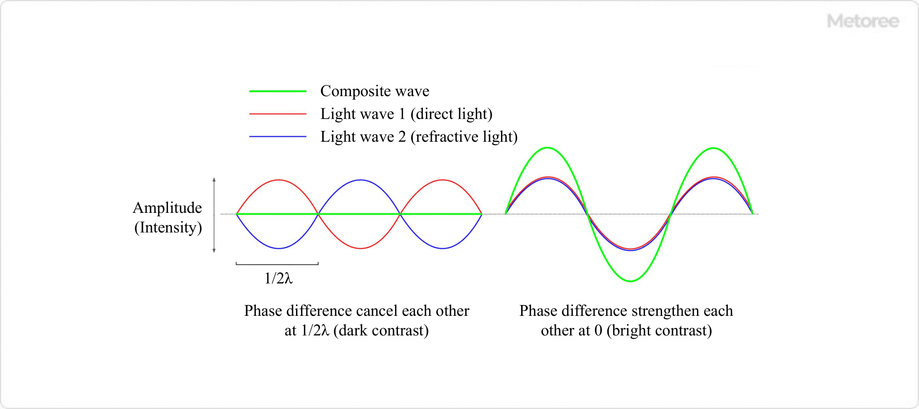
Figure 2. Interference between amplitude-matched direct light and diffracted light
In other words, at the site of a sudden change in refractive index where diffracted light is generated, when the phase difference is 1/2λ, direct light and diffracted light interfere with each other in a weakening manner, resulting in a dark appearance. This is the dark contrast. On the other hand, when the phase difference is 0, direct light and diffracted light interfere with each other in such a way that they strengthen each other, making the site of the abrupt change in refractive index appear bright. This is the bright contrast.
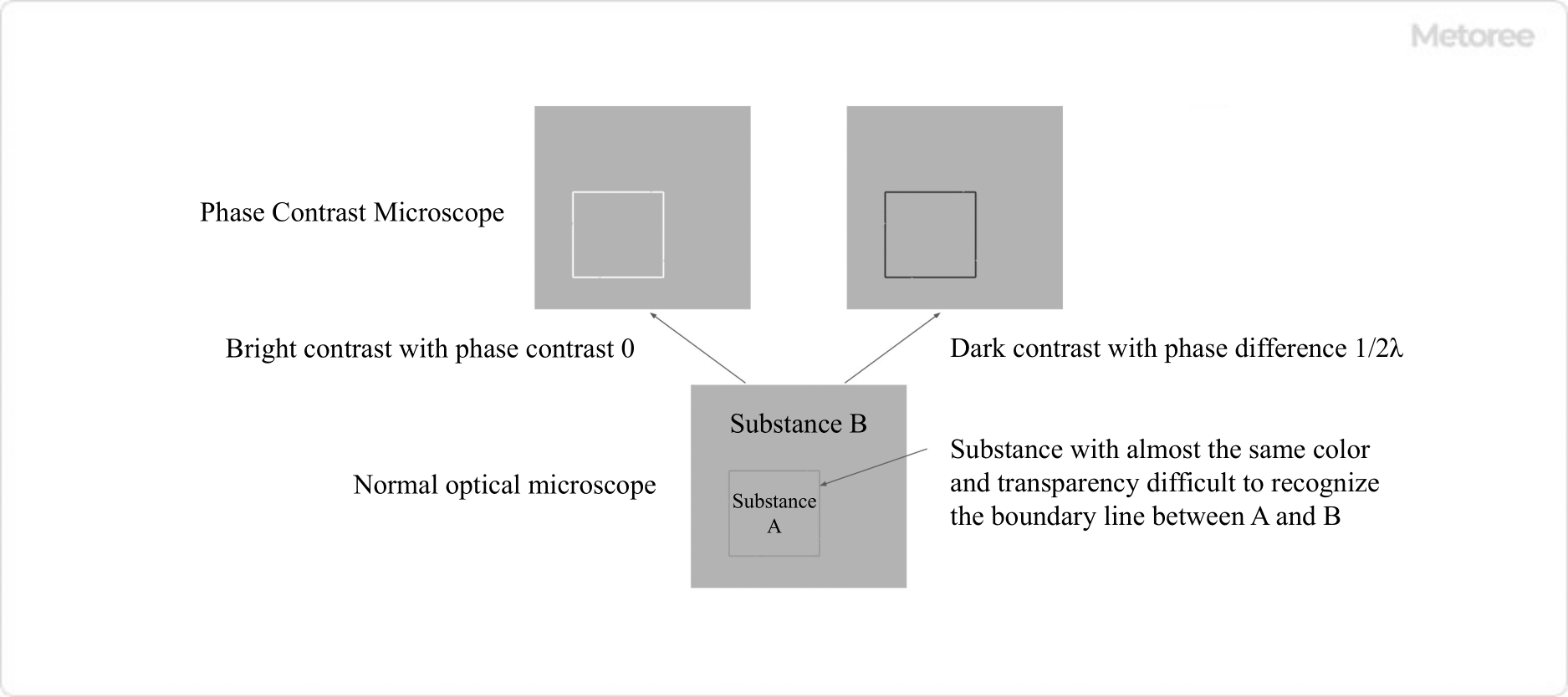
Figure 3. Bright contrast and dark contrast
Other Information on Phase Contrast Microscopes
1. Problems With Optical Microscopes
In ordinary optical microscopy, a substance can be identified by differences in either the intensity (amplitude) or the color (wavelength) or both of the light transmitted through the substance under observation. Therefore, for example, it is not easy to recognize the difference or boundary between a colorless transparent substance A and a colorless transparent substance B that is in contact with another colorless transparent substance A, even if they are observed with an ordinary optical microscope.
This is because there is no difference in the intensity and color of the transmitted light and no contrast between A and B. However, if the refractive indices of substances A and B are different, at the boundary between them, light is divided into direct light that travels straight through the sample and diffracted light whose path is altered. Since diffracted light is generated at the point where the refractive index changes abruptly, it contains information about the boundary shape and internal structure of each material in the sample.
It is important to note that diffracted light is delayed by a quarter of a wavelength (λ) (1/4λ) relative to the direct light traveling straight through the sample. Such a delay of a fraction of a wavelength is called a phase difference. Even if diffracted light is generated, the phase difference is minute because it is very weak compared to direct light.
Therefore, the image formed by adding the direct light and diffracted light together has a wave shape similar to that of the direct light, and no contrast between bright and dark is produced with an ordinary optical microscope.
2. Difference Between Phase Contrast Microscopes and Differential Interference Microscopes
In addition to phase contrast microscope, differential interference microscope is another type of microscope that uses light interference to obtain contrast. In differential interference microscopy, the light incident on a sample is separated into two polarizations with slightly different paths, and the two lights interfere with each other after passing through the object of observation to obtain contrast.
It is similar to the phase contrast microscope in that it can observe colorless and transparent materials, which is impossible with the phase contrast microscope. However, phase contrast microscope provides contrast in areas where the refractive index of the sample changes abruptly, whereas differential interference microscope provides contrast in areas where there is a gradient in the thickness or refractive index of the sample.