What Is a CT Scanner?
 A CT (Computed Tomography) Scanner is a scanner used for inspection.
There are two main types of CT Scanners: Medical CT Scanners used for imaging the human body and animals, and Industrial CT Scanners used for non-destructive testing and shipping inspection of products.
Uses of CT Scanners
CT scanners are used in the medical field to diagnose abnormalities in the brain and lungs.
In the industrial field, they are commonly used to detect the internal structure of products to measure the internal dimensions of wiring, to elucidate the internal state of forged products, to measure errors, and to detect the alignment of fibers.
When used in the medical field, X-ray output must be suppressed to avoid excessive exposure to radiation, but when used for industrial applications, X-ray output can be increased without any problem, thus enabling information to be obtained with higher precision than in medical applications.
Principle of CT Scanners
The principle of a CT scanner is similar to that of an X-ray. They consist of an X-ray irradiator, a detector, and an image processor that converts the detected data into an image.
Since each material absorbs X-rays differently (X-ray absorption coefficient), when X-rays are irradiated to an object to be examined, they penetrate at different rates of transmission depending on the material. Using this principle, we irradiate an object to be inspected with X-rays and detect the different transmission doses for each part of the object to be inspected. The general principle is to create an image by image processing based on this detection data.
The major difference between X-rays and CT scanners is that X-rays are irradiated from one direction on the specimen, while CT scanners irradiate X-rays from various directions on the specimen. For this reason, X-rays produce only flat images based on two-dimensional data, while a CT scanner uses three-dimensional data to produce a series of images of the specimen sliced in a circle at multiple locations, as well as images and movies that are composed in three dimensions.
Two physical phenomena are used in CT scanner produced scans: Compton scattering and the Photoelectric effect.
1. Compton Scattering
This occurs when X-rays collide with electrons, imparting some of the energy of the X-rays to the electrons, causing them to be blown out of their atomic orbits and reducing the energy of the X-rays.
2. Photoelectric Effect
This occurs when X-rays collide with electrons and all the energy of the X-rays is absorbed by the electrons, causing the electrons to be blown away from their atomic orbits and the X-rays to disappear.
Differences Between CT Scanners
There are two main types of CT Scanner applications: medical and industrial. The differences between them are explained from the following perspectives:
1. X-Ray Output
Medical CT scanners need to suppress X-ray output to avoid excessive exposure. On the other hand, industrial CT scanners can use relatively high X-ray output because the object to be inspected is an object, and thus can obtain highly accurate information.
2. Configuration
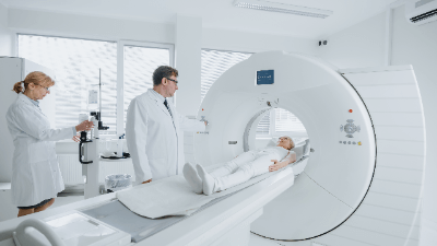
Medical CT Scanner
A medical CT scanner consists of a doughnut-shaped gantry in which an X-ray irradiator and a detector are placed relative to each other. The gantry has a bed-like part on which the object to be examined is placed within the ring of the gantry. The gantry rotates around the bed, and the X-ray irradiator and detector rotate around the object to be inspected.
Industrial CT Scanner
In the industrial CT scanner, the X-ray irradiator and detector are fixed relative to each other, and the part to be inspected is placed between them. By rotating the part on which the object to be inspected is placed, the object itself is rotated for inspection.
Therefore, depending on the size of the object to be inspected, industrial CT scanners can be made smaller.
Industrial CT scanners are equipped with an X-ray leakage cabinet to confine X-rays inside the device, which also makes it possible to reduce the size of the scanner.
In both medical and industrial CT scanners, there are two types: one with a single row of detectors along the X-ray transmission plane in the width direction of the object to be inspected, and the other with multiple rows of detectors along the length direction of the object to be inspected.
The one with multiple rows of detectors can acquire data for each row at a time, which makes it possible to acquire data faster than the one with only one row.
In the case of industrial CT scanners, multiple-row CT Scanners are more suitable because efficiency is more important. Medical CT scanners are also often used because the faster the examination can be completed, the less burden is placed on the patient.
Other Information on CT Scanners
1. The Relationship Between CT Scanners and Cancer
CT scans are widely used for everything from examination to diagnosis and even medical treatment, although the image of CT scans is that they are used to find cancer. Although, there is a risk of cancer from CT scans, and it is necessary to fully understand the risks when CT scans are performed.
A CT scanner is an important device in medicine, but it is not without risks of causing cancer because of the x-ray irradiation.
The risk of developing cancer from a single CT scan is considered low, and the risk of not undergoing a CT scan is much higher than the risk of undergoing a CT scan.
2. Difference Between CT and MRI
The main difference between CT and MRI is the principle of measurement.
CT scanners form images based on differences in the transmission of X-rays through different materials, as described above, whereas MRI creates images based on the magnetic resonance of materials. CT scanners have the advantage of short acquisition times and the ease of obtaining tomographic images, and are more indicated for emergency head lesions (such as suspected hemorrhage). and the ability to image bone.
On the other hand, MRI is inferior to CT in terms of radiation exposure and differences in density between lesions and normal tissue.
Another advantage is that any tomographic image can be obtained, and images of blood vessels can be obtained without the use of contrast media. The disadvantage, on the other hand, is that those with implanted devices in the body cannot be examined.