What Is a Laser Microscope?
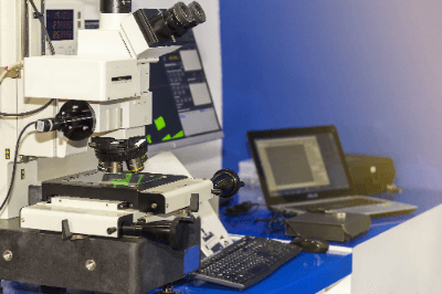 A laser microscope is a type of optical microscope in which a sample can be observed by scanning a laser beam to a light source.
It generally employs confocal optics and is also called a confocal laser scanning microscope or CLSM. A laser microscope has high spatial resolution not only in the horizontal (XY) direction but also in the vertical (Z) direction because the confocal optics can exclude light from out-of-focus surfaces.
Therefore, by measuring microscope images while shifting them in the height direction, it is possible to obtain a three-dimensional image or an all-in-focus image.
Uses of Laser Microscopes
Since laser microscopes use light for measurement, there is no need to touch the sample. For this reason, laser microscopes are used in the industrial field to observe the three-dimensional shapes and surface profiles of precision instruments, such as semiconductors and electronic components. They are also used in the life science field to observe cells and biological tissues labeled with fluorescent substances.
Some manufacturers offer customized measurement stages for laser microscopes, making it possible to measure large samples such as large flat panel displays.
Principle of Laser Microscopes
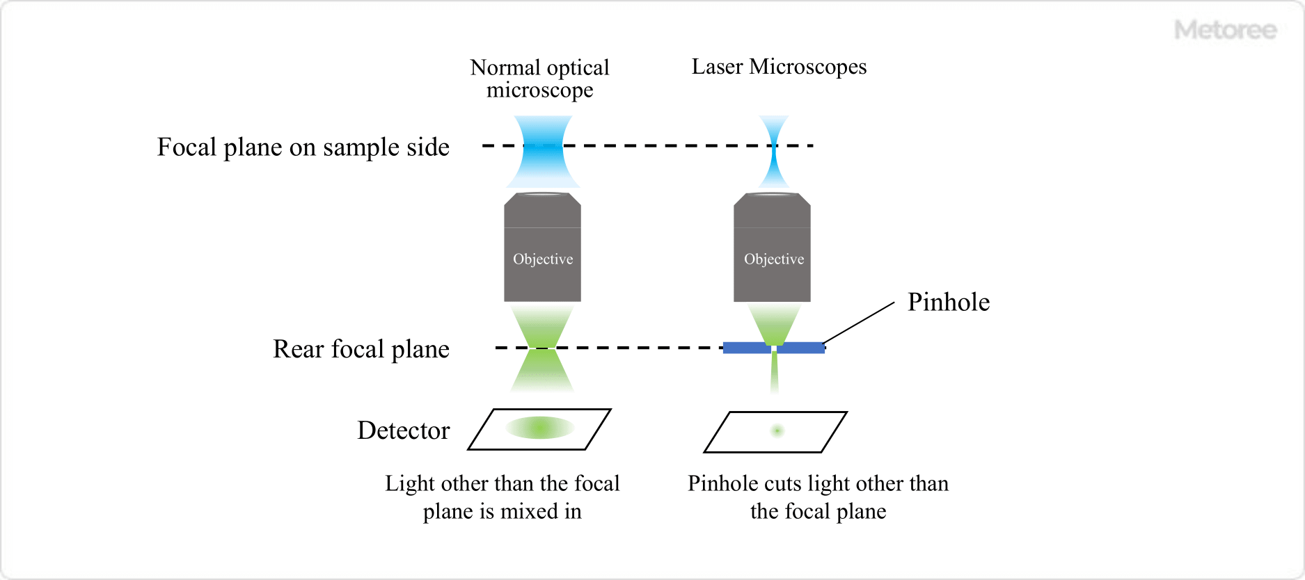
Figure 1. Comparison of optical and laser microscopes
Laser microscopes are similar to a typical optical microscope configuration of lenses and mirrors, but use a laser as the light source and are designed as a confocal optical system. Laser light is characterized by the uniform wavelength and phase of the emitted light, and by its excellent monochromaticity, directivity, and linearity.
Since ordinary light has different phases and wavelengths, the optical paths are not aligned, and the reflected light generated by irradiating a sample is overlapped by scattered light, making it difficult to obtain a clear image. On the other hand, with laser microscopes, a pinhole is placed at the position where the reflected light is focused through transmission through the lens and reflection from the sample. Therefore, excess light, such as scattered light, can be eliminated. As a result, clean images with clear contours can be obtained.
In addition, there are two methods of obtaining two-dimensional images with laser microscopes: one is to move the stage, and the other is to move the laser mechanism. The features of each method are as follows.
- The Stage-Moving Method
A wide area can be measured, but the size of the stage is limited, so large samples cannot be measured. - Laser MechanismMoving Method
A wide range of sample sizes can be measured, and surface microstructures can also be measured.
Scanning Method of Laser Microscopes
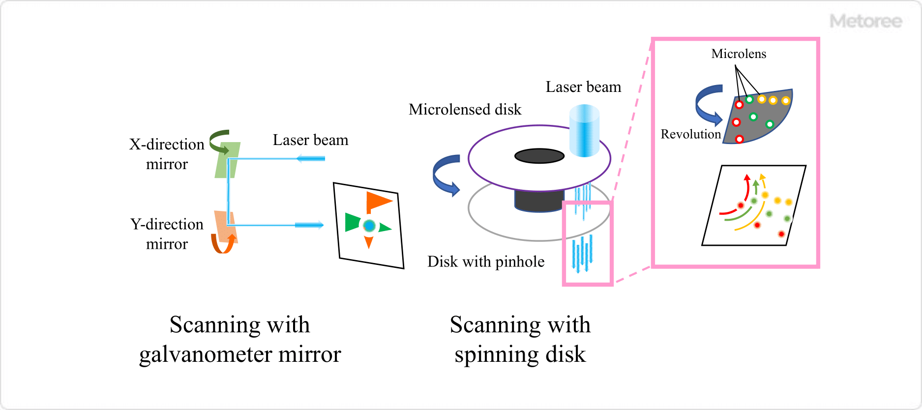
Figure 2. Laser microscope scanning method
There are various ways to scan with laser microscopes. For example, scanning with a galvano mirror involves mechanically moving the mirror, but a MEMS scanner or resonant scanner method can be used to increase the speed.
The spinning disk method is used for high-speed measurement, in which a laser beam is directed onto a disk lined with many micro-lenses and pinhole arrays to pick up many beams of light simultaneously reflected from the sample. This method can obtain a large amount of information at a time but requires a high-power laser that has sufficient intensity even when spread to some extent.
Other Information About Laser Microscopes
1. Differences Between Laser Microscopes and Electron Microscopes
The electron microscope is another type of microscope with high magnification, but the principles of these instruments are not identical. Laser microscopes use light, while electron microscopes use electron beams, and the magnification, equipment, and measurement techniques are very different.
Because electrons are very short in wavelength compared to visible light, the resolution of electron microscopy is very high; scanning electron microscopes (SEM) can observe structures down to a few nanometers. Laser microscopes, on the other hand, cannot observe structures at wavelengths shorter than their wavelengths and have a resolution of only a few hundred nanometers.
The equipment used for laser microscopy and electron microscopy differs greatly. Electron microscopy uses an electron beam and requires measurement under a vacuum. In addition, when highly insulating materials are measured with electron microscopes, the electron beam may cause charge to accumulate on the surface, distorting the image, and other limitations exist, so care must be taken to determine what the sample’s unique physical properties are.
Also, as a measurement technique, electron microscopy requires skillful techniques for cutting out the surface and optimizing the observation conditions. On the other hand, laser microscopes can be used more universally than electron microscopes because there is no accumulation of electric charge and the surface cutout does not require precision.
2. Surface Roughness Measurement With Laser Microscopes
Confocal laser microscopes can measure the roughness of a sample surface in a non-contact manner. Although atomic force microscopy is the most common method for measuring the roughness of a sample surface, confocal laser microscopy has the advantage of non-contact measurement. Conversely, the resolution differs from that of the atomic force microscope, so the appropriate instrument should be selected depending on the roughness of the sample surface.