What Is a Stereo Microscope?
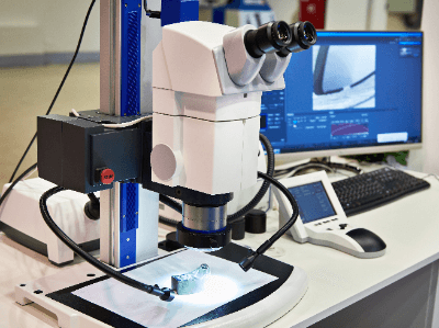
A stereo microscope is a type of optical microscope, also called a binocular stereo microscope. An optical microscope is an optical instrument in which the actual image of a microscopic object magnified by an objective lens is further magnified by an eyepiece lens for observation. Stereo microscopes have two light paths, and by using the parallax between the left and right eyes, objects can be observed in three dimensions. In general, the magnification is relatively low, ranging from several times to 40 times, but the long distance between the objective lens and the stage makes it possible to observe relatively large specimens as they are. In addition, dissection, assembly, and other operations can be performed while viewing the magnified image.
Types and Features of Optical Microscopes
There are various types of optical microscopes depending on the principle of and purpose for which they are used. They are generally classified into two types: biological microscopes and stereo microscopes. Biological microscopes are a type of transmission microscope and should be considered the most common type of microscope. This type of microscope observes a sample by making a thin slide of the sample and allowing light to pass through it.
Stereo microscopes, on the other hand, use both transmitted and oblique illumination. Stereo microscopes have two eyepieces that allow simultaneous observation with both eyes, making it possible to observe the specimen in three dimensions. In addition, the long distance between the eyepieces and the stage on which the specimen is placed makes it suitable for tasks such as dissecting a biological specimen while observing it.
How to Use a Stereo Microscope
The general instructions for using a stereo microscope are as follows.
- Set the light source as necessary.
- Place the specimen on the stage.
- Adjust the eyepiece to fit the width of the eye so that the fields of view overlap into one.
- Focus the sample by looking through the right eyepiece with the right eye and operating the focusing device.
- While looking through the left eyepiece with the left eye, adjust the diopter adjustment ring to bring the sample into precise focus.
Uses of Stereo Microscopes
In the biological field, stereomicroscopes are used for observation and dissection of animals and plants, such as insects and flowers. They are also used as science teaching materials in educational settings. In the medical field, they are used for dissection and cell manipulation. The microscopes used in neurosurgery and ophthalmology during surgery are also stereo microscopes. In the fields of mechanical engineering, precision machinery, and electronics, microscopes are used for assembly and inspection. Stereo microscopes are also used in a wide range of other applications, such as in dental work, handicrafts, soldering, and other precision work, as well as in the observation and study of old coins and jewelry.
Principle of Stereo Microscopes
There are two types of lenses: “positive lenses” (convex lenses) and “negative lenses” (concave lenses). Positive lenses are used in microscopes.
The center of a positive lens is thicker than its edges, and it refracts light parallel to a line (optical axis) perpendicular to the lens through the center of the lens surface and collects it at a point on the optical axis. This point is called the “focal point.”
A positive lens has one focal point at the front and one at the rear of the lens (front focal point and rear focal point), and the distance between the focal point and the center of the lens is called the “focal length”. The focal lengths of the front and rear foci are equal. The real image formed by the lens when an object is farther from the front focal point of the positive lens is called the “real image” and the image formed when the object is closer to the front focal point is called the “false image.” The real image is an inverted image with the vertical and horizontal sides reversed, while the imaginary image is an upright image.
A typical biological microscope is a device that combines two positive lenses to magnify an object. This device observes the real image formed by the objective lens (lens close to the object) and the imaginary image further magnified by the eyepiece lens (lens close to the eye), so the observed image is inverted.
Stereo microscopes, on the other hand, have an upright prism built into the body, so the observed image is upright. This allows the sample to be observed as it is and enables precise work under the microscope.
Features and Types of Stereo Microscopes
Stereo microscopes are available in a variety of models with different specifications, which should be selected according to the use of the microscope. The following differences, for example, are important points to consider when selecting a microscope.
Difference in Optical System
Stereo microscopes are broadly classified into two types: Galilean parallel optics and Grineau optics. Microscopes with Galilean parallel optics consist of a single objective lens with a parallel optical axis from the eyepiece to the objective lens. Since the optical axis is designed to be parallel, it is possible to add various functions by inserting another unit in the middle.
In addition, since they converge light on a single objective lens, observation at high magnification is possible. Accuracy is easily maintained even when the zoom is increased, and there is a high degree of freedom in the combination of objective lenses. On the other hand, microscopes with Grineau-type optics are designed so that the optical path and optical axis from the eyepiece to the objective lens are all independent of left and right at a certain angle. This is a feature that makes it easy to obtain a three-dimensional image and to design a compact microscope body. However, since there is no parallel part of the optical path, it is not suitable for adding another function to the middle part of the optical path or for setting a large zoom range, as is the case with Galilean lenses.
Difference in Illumination
In stereo microscopes, the choice of illumination is also important for optimum observation of the specimen. Illumination should be selected to match the microscope used and the purpose of the observation. Types of illumination include ring light for bright and uniform illumination, near vertical illumination for fewer shadows, and coaxial illumination for observation of flat specimens with high light reflectance. Halogen lamps and LEDs are generally used as light sources.
Magnification of Stereo Microscopes
There are three types of microscope magnifications: objective magnification, total magnification, and monitor magnification. Objective magnification refers to the magnification of the objective lens alone. Total magnification is the product of the magnification of the objective lens and the magnification of the eyepiece. In a microscope, the image obtained by the objective lens is magnified by the eyepiece, so even if the image has the same overall magnification, the higher magnification of the objective lens has a higher resolution and enables the determination of finer points. Monitor magnification refers to the magnification of an image when it is displayed on a monitor display and indicates how many times larger the image appears when projected on a monitor. The same magnification number can be viewed differently depending on what the magnification represents.