What Is a Transmission Electron Microscope?
A transmission electron microscope (TEM) is a measurement device that can observe the internal structure of a sample.
It is a type of electron microscope that observes the inside of an ultra-thin sample by irradiating electron beams onto the sample and detecting the transmitted and scattered electrons that pass through the sample. The technique is used in various fields, including materials engineering and biochemistry, as it enables the observation of the internal structure of a sample at a high magnification, which is not feasible with an optical microscope.
Uses of Transmission Electron Microscopes
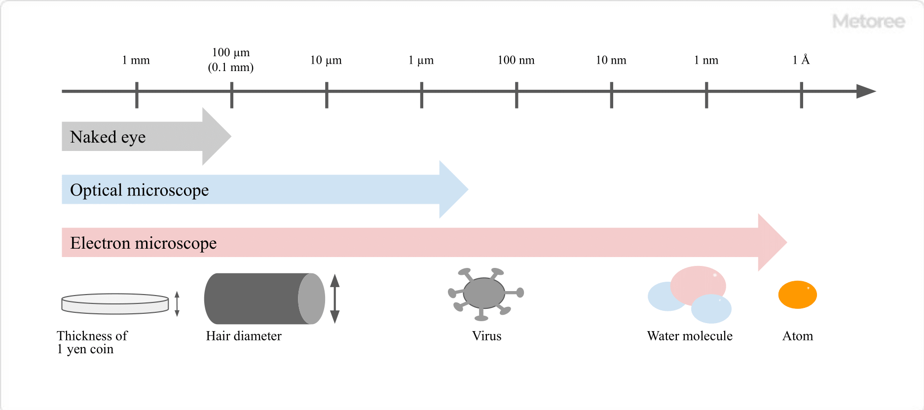
Figure 1. Types and resolutions of microscopes
TEMs are used to observe the internal structure of a sample at magnifications of several hundred to several million times.
It is possible to observe entire cells at the level of tens of micrometers, as well as atomic structures of atomic arrangements at the level of a few Å (1Å (angstrom) = 10-10m). It finds application in the structural analysis of diverse materials, such as semiconductors and ceramics, as well as in the structural analysis of biological samples like cells and bacteria. Various information can be obtained, such as observation of electron diffraction patterns by adjusting the lens system, and elemental analysis and state analysis by attaching an additional spectrometer. Unlike scanning transmission electron microscopes (STEM), TEMs are sometimes used to observe changes in the structure of an object over time, since image data can be acquired at once.
Principle of Transmission Electron Microscopes
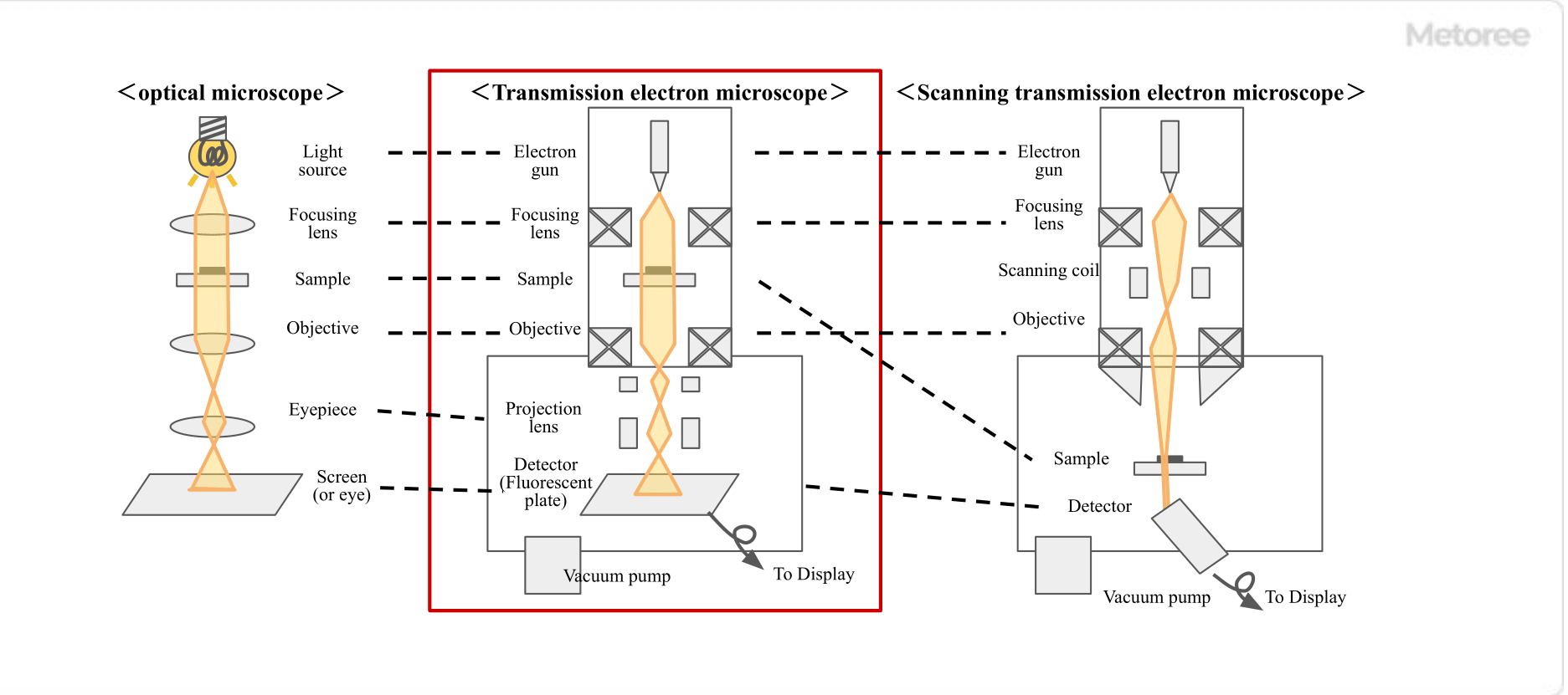
Figure 2. Types of microscopes and structural images
The principle of TEMs is to irradiate a sample with accelerated electrons and observe its internal state by detecting the electrons transmitted through the sample. The structure of the microscope is similar to that of an optical microscope. However, since the light source is an electron beam rather than visible light, the thickness of the sample must be reduced to a level where electrons can penetrate (100 nm or less). The difference in the density of electrons transmitted through the sample appears as a contrast.
The shorter the wavelength of the electrons irradiating the sample (the higher the energy), the higher the resolution (~0.1nm), since the wavelength of electrons accelerated at an acceleration voltage of 300kV is 0.00197nm, which is much shorter than the wavelength of visible light (about 380nm to 780nm) used in optical microscopy.
The higher the acceleration voltage, the shorter the wavelength and the higher the resolution. However, this increases the damage to the sample and must be adjusted appropriately. The upper limit of resolution is about 50pm due to factors such as aberration of the optical system.
Other Information on Transmission Electron Microscopes
1. Preparation of Samples for Transmission Electron Microscopes
Some samples require appropriate sample preparation.
Thick Specimens
Samples to be observed by general transmission electron microscopes need to be thinned to a thickness of about 100nm.
1. Dispersion Method
The sample is dispersed in a solvent, and the dispersion is dropped onto a substrate for observation.
2. Microtome Method
This method uses a diamond knife to thin the sample to about 100nm. Soft samples such as polymers are cooled with liquid nitrogen and then cut.
3. Ar Milling Method
A sample that has been mechanically thinned to a thickness of several tens of micrometers is irradiated with Ar+ ions, which breaks the bonds in the sample while thinning it.
4. FIB Method
The target area is thinned by FIB while observing with a scanning electron microscope (SEM). Using an ultra-high voltage electron microscope (HVEM) with an acceleration voltage of 1000kV or higher, it is possible to observe samples with a thickness of about 5µm. However, since the equipment is very large and the structure of the microscope is complex, it is mainly owned by research facilities such as universities.
Samples That Do Not Contain Heavy Elements
Polymer and biological samples are mainly composed of light elements such as C, H, N, and O, which are highly permeable to electrons and may not provide sufficient contrast to identify the Structure of the sample. Selective electron staining with a staining agent with high electron scattering capacity (e.g., OsO4 or RuO4) in the area where the structure of the sample is to be observed will provide an image with sufficient contrast. Electron staining can alter the Structure of the sample. To avoid this effect, it is effective to use the phase contrast method of transmission electron microscopy or scanning transmission electron microscopy (STEM).
Samples That Evaporate or Sublimate Under High Vacuum Conditions
Evaporation or sublimation under high vacuum conditions not only changes the Structure of the sample and its shape but can also lead to equipment failure. To prevent this, environmentally controlled transmission electron microscopes (ETEM) or cryo-electron microscopes should be used.
2. The Main Analytical Equipment Attached to Transmission Electron Microscopes>
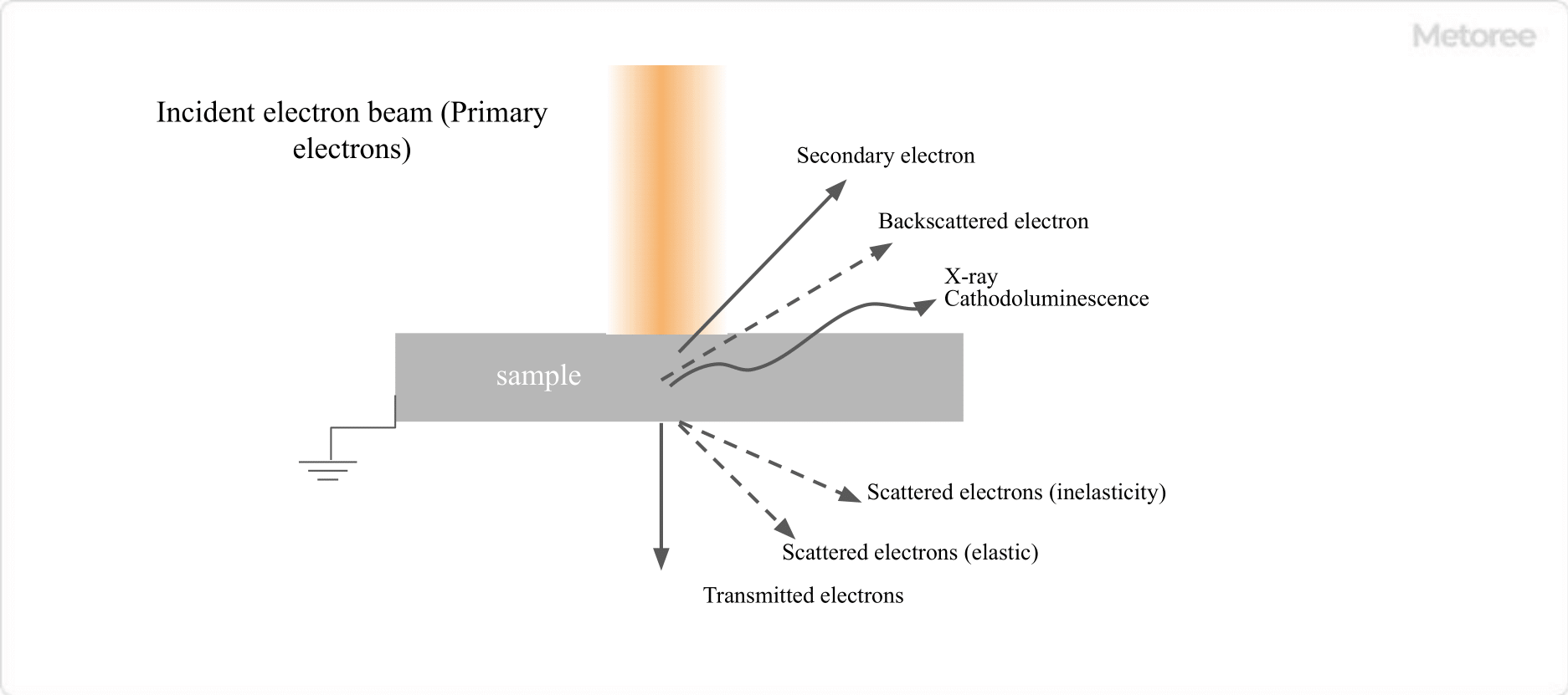
Figure 3. Main electromagnetic waves generated by electron beam irradiation
Since various signals other than electrons can be obtained by irradiating a sample with an accelerated electron beam, transmission electron microscopes may be equipped with various types of analytical instruments.
Electron Beam Diffraction
By detecting the interference of elastically scattered electron beams, a diffraction image of the sample is obtained. Analysis of the diffraction image reveals crystallographic information, such as the Structure of the crystal and its orientation.
Electron Energy Loss Spectroscopy (EELS)
Inelastically scattered electron beams are those emitted from a sample after the incident electron beam excites electrons in the sample. By measuring how much energy is lost by the electron beam compared to before the incident beam, information about the composition and bonding state of the sample can be determined.
Electron Tomography
By applying the principles of CT (computer tomography) to transmitted electrons, we can produce a three-dimensional stereoscopic image of a sample by stacking cross-sectional images of the sample.
Various other analytical functions can be added to these. Compared to measurements made with an independent measuring device, more detailed measurements can be made because the measurement position can be selected while viewing the transmission electron microscope image.